Nowoczesna protetyka stomatologicznej jest dziedziną zdigitalizowaną. Protetyka cyfrowa oznacza zastosowanie technologii cyfrowych praktycznie na każdym etapie leczenia protetycznego, począwszy od cyfrowych zdjęć 3D, umożliwiających ocenę stanu zębów i ich położenia w kości, przez cyfrową analizę okluzji, po projektowanie i drukowanie uzupełnień protetycznych. Jednak cyfrowo nie zawsze oznacza bezbłędnie.
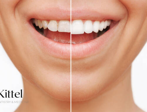
Protetyka stomatologiczna to dział stomatologii, który zajmuje się odtwarzaniem prawidłowego zgryzu za pomocą uzupełnień stałych: koron, licówek i mostów lub ruchomych – protez. Protetyk stomatologiczny pomaga również w sytuacjach, kiedy pacjenci chcą po prostu nadać swoim zębom bardziej estetyczny wygląd.
Protetyka cyfrowa XXI wieku
Współczesne leczenie protetyczne opiera się na podejściu holistycznym. Oznacza to, że podłożem protetycznym, czyli punktem odniesienia w pracy protetyka, jest cały układ stomatognatyczny pacjenta, a nie pojedynczy ubytek. Odtwarzając ząb lekarz bierze więc pod uwagę nie tylko stan i położenie pozostałych zębów i to jak ze sobą kontaktują, ale również morfologię twarzy, tor oddechowy czy położenie kręgosłupa. Co więcej, podłoże to musi być odpowiednio przygotowane. Oznacza to wygojone dziąsła, wyleczone z próchnicy i stanów zapalnych oraz oczyszczone z kamienia zęby.
Drugą istotną cechą nowoczesnej protetyki stomatologicznej jest jej digitalizacja. Techniki cyfrowe są stosowane praktycznie na każdym etapie leczenia protetycznego. Zaczynamy od cyfrowych zdjęć 3D, umożliwiających ocenę stanu zębów i ich położenia w kości. Następnie cyfrowo analizujemy okluzję, a na końcu projektujemy i drukujemy uzupełnienia protetyczne. Nowoczesne gabinety stomatologiczne i laboratoria protetyczne pracują w pełni zdigitalizowany sposób. Jednak nie we wszystkim cyfryzacja wyręczy lekarza i technika dentystycznego, o czym napiszę w dalszej części tekstu.
Digital prosthetics – from the analysis of occlusion to the placement of restorations in the patient's oral cavity
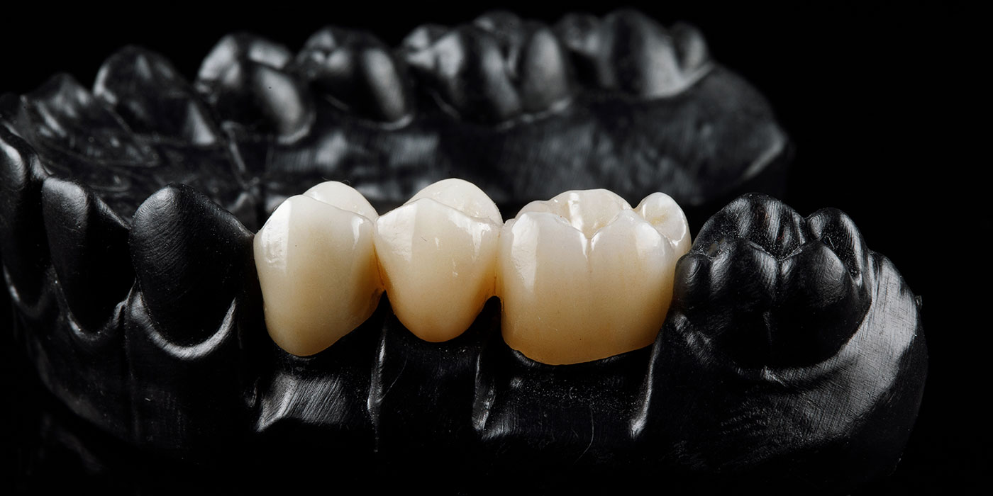
Najczęściej wykonywanymi uzupełnieniami protetycznymi są korony protetyczne. Są one osadzane pojedynczo lub w formie mostów na naturalnych korzeniach zębów bądź implantach. W klinice Dr Kittel uzupełnieniem pierwszego wyboru są pełnokonturowe korony cyfrowe wykonywane z tlenku cyrkonu, o których więcej pisałam tutaj. Dzięki eliminacji nietransparentnego metalu z podbudowy stosowanej w koronach porcelanowych, ten rodzaj uzupełnienia gwarantuje estetykę na najwyższym poziomie. Jakość koron protetycznych ma niebagatelne znaczenie dla sukcesu całego leczenia, bo to właśnie korony pełnią niejako funkcję ortezy dla stawów skroniowo-żuchwowych. Zęby są tu tylko podparciem. Na przykładzie koron „full contour” omówię jak przebiega cyfrowe leczenie protetyczne.
1. Digital prosthetic treatment planning
The first stage of a prosthodontist's work is to recognize the problem and make a diagnosis. Already at this stage, in addition to the clinical examination, the doctor has digital tools at his disposal, such as digital radiological examinations and an intraoral scanner. In most cases, the scanner replaces existing analog (plaster) impressions. The device uses a beam of light that transfers the image of hard and soft tissues from the patient's oral cavity to a computer language. The image obtained in this way can be enlarged and rotated. The undoubted advantage of digital scanning is its accuracy, which remains unchanged throughout the entire work process in the office and prosthetic laboratory. However, digital does not always mean error-free. It turns out that we cannot rely 100 percent on technology in all cases. In the case of large, full-arch works or complete edentulism (dentures), it is better to rely on traditional impressions, as the scanner may have difficulties in correctly registering all fragments of the mucous membrane.
2. Digital smile visualization
Digital technology also allows us to pre-design the smile and visualize the patient's face after treatment, i.e. plan the aesthetics before the treatment begins. Virtual design takes into account both the patient's expectations and the existing occlusal conditions. Visualization of the final effect of treatment at this stage allows for its proper planning (including, for example, determining whether orthodontic treatment is justified).
3. Digital occlusion analysis and design (CAD phase)
Oczekiwana estetyka zębów zawsze musi iść w parze z prawidłową funkcjonalnością. Wiedząc już jaki efekt chcemy osiągnąć i jakim sposobem, przystępujemy do cyfrowej weryfikacji okluzji. Okluzja to inaczej kontakty pomiędzy górnymi i dolnymi zębami. Nieprawidłowa okluzja (zwarcie zębów) może skutkować bólem, a nawet problemami z żuciem pokarmów i mówieniem. Dlatego przy odbudowie protetycznej kluczowe jest jej właściwe zaprojektowanie. Z pomocą przychodzi tu specjalne oprogramowanie, do którego trafiają pliki z wykonanymi wcześniej cyfrowo skanami wewnątrzustnymi. Projektowanie obejmuje m.in. ustalenie kąta osadzenia przyszłych koron na zębach, które będą ich filarami i wyznaczenie zasięgu granic preparacji, czyli oszlifowania zębów. Wykonany w ten sposób projekt jest analizowany w wirtualnym artykulatorze. Działa on na podobnych zasadach do artykulator manualny. Symulacja odwzorowuje naturalne ruchy żuchwy względem szczęki, w tym ruchy boczne, doprzednie, tzw. prowadzenie kłowe czy sieczne. Zwarcie można dowolnie podnieść lub obniżyć dostosowując je do warunków anatomicznych pacjenta. Wszystko po to, by przyszłe korony nie powodowały dyskomfortu czy dysfunkcji w stawach skroniowo-żuchwowych oraz nie wymagały znaczących korekt po ich wydrukowaniu.
Based on the initial virtual design, the so-called mockup – an individual 3D design placed on the patient's teeth or "virtually tried on". Analog mock-up allows you to "try on" new teeth at a stage when it is still possible to make changes to the design. After the patient's acceptance, the doctor starts preparing the teeth for future crowns, and the laboratory starts working on the final restorations.
4. Preparation of teeth for crowns
Preparation of teeth for crowns involves preparing them for permanent placement of prosthetic restorations. The teeth must be properly ground to provide support for the crowns. It includes the incisal edges, occlusal surface, proximal, labial, palatal and cervical surfaces. Modern digital technologies that we use when designing crowns mean that tooth grinding is reduced to a minimum. The preparation should be carried out in a way that ensures perfect marginal tightness and good adhesion of the crown.
5. Crown milling (CAM phase)
In the prosthetic laboratory, the individual, digital design of the crowns goes to a milling machine that cuts the restorations from special ceramic blocks. Modern CAM technologies enable machine-made crowns with an appropriate texture, occlusal surface and excellent marginal tightness. After milling, the crowns are ready for characterization, which is done manually. Dental technicians work to ensure that the appearance of crowns is as close to natural as possible, imitating the natural dynamics of light by selecting appropriate paints and glazes. At this stage, artificial intelligence cannot replace human artistry.
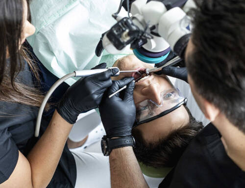
6. Cementation of crowns
Ready-made prosthetic crowns are sent from the laboratory to the office, where the prosthodontist cements (i.e. permanently embeds) them on pillars (prepared teeth) or on implants. The entire process ends with digital and analog occlusion verification, and necessary corrections are made if necessary.
The use of digital techniques for analysis, design and production of prosthetic works allows for a radical shortening of the treatment process and makes it more predictable and precise. In the past, we somehow adapted our prosthetic work to occlusal conditions, but today we can influence these conditions. Teeth grinding, i.e. interference with their living structure, has been limited to a minimum. The precision of the design and printing of crowns affects the comfort and durability of their use. The technology uses an image that is understandable to the patient. Finally, technology standardizes processes and significantly reduces the risk of human errors, although, as I mentioned earlier, it can make mistakes itself, which is why the participation of a doctor and a dental technician is necessary in the entire process.
Leave A Comment